Search results
Search for "binding energies" in Full Text gives 156 result(s) in Beilstein Journal of Nanotechnology.
Investigating ripple pattern formation and damage profiles in Si and Ge induced by 100 keV Ar+ ion beam: a comparative study
Beilstein J. Nanotechnol. 2024, 15, 367–375, doi:10.3762/bjnano.15.33

- and energy of the incoming ion and on the mass of the target atom. It may be expressed as the spatial distribution of the energy transferred/deposited within the target [27][28]. Sometimes the energy distribution on the target atoms at the surface may be sufficient to overcome binding energies so as


Controllable physicochemical properties of WOx thin films grown under glancing angle
Beilstein J. Nanotechnol. 2024, 15, 350–359, doi:10.3762/bjnano.15.31

- curve fitting after Shirley background subtraction [38]. The two major symmetric peaks at 35.87 and 38.00 eV binding energies correspond to the 4f7/2 and 4f5/2 levels (spin–orbit splitting: 2.13 eV), respectively, indicating the presence of W6+ in the as-deposited WOx films [38][39]. The two minor peaks
- at 34.78 and 36.92 eV can be designated to 4f7/2 and 4f5/2 levels of the W5+ oxidation state [40]. Similarly, the presence of W6+ and W5+ is observed in the annealed WOx films, where the W 4f7/2 and W 4f5/2 peaks corresponding to the W6+ state are found at slightly smaller binding energies (35.82 and


Investigating structural and electronic properties of neutral zinc clusters: a G0W0 and G0W0Г0(1) benchmark
Beilstein J. Nanotechnol. 2024, 15, 310–316, doi:10.3762/bjnano.15.28

- ionization potential compared to other methods. Keywords: binding energies; CALYPSO structure prediction; DFT; G0W0 studies; zinc clusters; zinc isomers; Introduction Zinc is a group-IIB element that is frequently used as a galvanizing material and in storage media as an anode [1][2][3]. However, its
- presented, and binding energies are discussed. In addition, we have also explored the ionization potentials, electron affinities, and energy gaps for the series of Zn clusters. Geometrical structures Various theoretical studies have been performed regarding the structural properties of Zn clusters. Among
- such, a DFT study employing the PBE functional revealed that the symmetric structures are less stable than the structures with lower symmetry [6]. There are also experimental and theoretical studies to determine the binding energies of Zn clusters [4][5][25]. In addition, in an experimental study also


![[Graphic 14]](/bjnano/content/inline/2190-4286-15-28-i15.svg?max-width=637&scale=1.18182)
Multiscale modelling of biomolecular corona formation on metallic surfaces
Beilstein J. Nanotechnol. 2024, 15, 215–229, doi:10.3762/bjnano.15.21

- surface. At the largest scale, our methodology employs a coarse-grained (CG) kinetic Monte Carlo (KMC) method [16] to simulate competitive adsorption of biomolecules onto the aluminum surface. To achieve this, we evaluate individual binding energies at various orientations (represented by heatmaps) for
- orientation of each individual protein, a primary coarse-graining step was performed. In this part, we use the UA model to predict the protein–NP binding energies. This model takes into account various factors, such as the material’s chemical composition, size, shape, surface roughness, charge
- results and detailed information on the calculation can be found in Supporting Information File 1, Figure S2 and Figure S3, which illustrate the variations in adsorption energies as a function of NP size. Within the range of 2–20 nm the binding energies of ALAC, BLAC, BC, and BSA show an initial increase


Low temperature atomic layer deposition of cobalt using dicobalt hexacarbonyl-1-heptyne as precursor
Beilstein J. Nanotechnol. 2023, 14, 951–963, doi:10.3762/bjnano.14.78

- the carbonyl groups [33]. No evidence could be found of cobalt carbide formation, which would result in binding energies of approximately 284 eV or below [34][35]. The oxygen 1s peak has its maximum at 532.2 eV (Figure 5). However, assuming just one feature will result in a poor fitting result with an


Titania nanoparticles for photocatalytic degradation of ethanol under simulated solar light
Beilstein J. Nanotechnol. 2023, 14, 616–630, doi:10.3762/bjnano.14.51

- are shown in Figure 4d and were fitted with two peaks. The peaks at binding energies of 529.9 and 530.5 eV are attributed, respectively, to oxygen bound to Ti4+ and the adsorption of –OH on the surface [50][51]. The XPS results do not show differences between the two series of samples and do not


Mixed oxides with corundum-type structure obtained from recycling can seals as paint pigments: color stability
Beilstein J. Nanotechnol. 2023, 14, 467–477, doi:10.3762/bjnano.14.37

- of end states that will be seen in the photoelectron spectrum as a multipeak envelope [25][26]. The Fe 2p spectrum (Figure 4e) shows the characteristic doublet of Fe 2p1/2 and Fe 2p3/2 at binding energies of 725.1 and 711.1 eV, respectively. The prominent peaks of Fe 2p are accompanied by
- characteristic Fe3+ satellites at higher binding energies (shifted by ca. 9 eV from Fe 2p3/2) [27], similar to that reported in [28]. Electronic spectroscopy In the UV–vis absorption spectrum of sample 1 (Figure 5a), the prominent band with a maximum centered at 588 nm can be attributed to the 4A2→4T2 transition


Evaluation of electrosynthesized reduced graphene oxide–Ni/Fe/Co-based (oxy)hydroxide catalysts towards the oxygen evolution reaction
Beilstein J. Nanotechnol. 2023, 14, 420–433, doi:10.3762/bjnano.14.34
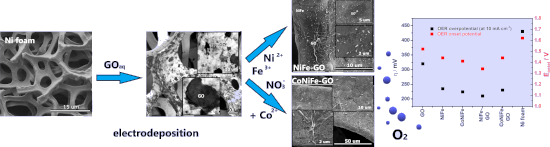
- calculations were determined based on the survey spectra collected in a wide range of binding energies, while valence state calculations were based on the high-resolution spectra. The C 1s peak (285.0 eV) was used to correct the results. Analysis of XPS spectra was performed with the Casa-XPS software using a


Spindle-like MIL101(Fe) decorated with Bi2O3 nanoparticles for enhanced degradation of chlortetracycline under visible-light irradiation
Beilstein J. Nanotechnol. 2022, 13, 1038–1050, doi:10.3762/bjnano.13.91

- increased electron density on Fe3+ [42][57]. As shown in Figure 4c, the Bi 4f spectra of BOM-20 were fitted by two peaks at 159.5 and 164.9 eV, which are assigned to Bi 4f7/2 and Bi 4f5/2, respectively, corroborating the presence of Bi3+ [52][57]. The binding energies of Bi 4f at 159.5 and 164.9 eV of BOM


Electrocatalytic oxygen reduction activity of AgCoCu oxides on reduced graphene oxide in alkaline media
Beilstein J. Nanotechnol. 2022, 13, 1020–1029, doi:10.3762/bjnano.13.89

- satellite peaks at 802.2 and 785.1 eV (Figure 6b). The peak difference between 2p1/2 and 2p3/2 and the satellite peak assignment represent the existence of cobalt atoms in Co2+ and Co3+ chemical states [34]. Likewise, the high-resolution spectrum of copper displayed Cu 2p1/2 and Cu 2p3/2 binding energies at


Hierarchical Bi2WO6/TiO2-nanotube composites derived from natural cellulose for visible-light photocatalytic treatment of pollutants
Beilstein J. Nanotechnol. 2022, 13, 745–762, doi:10.3762/bjnano.13.66

- , and W. The high-resolution XPS spectrum of the Bi 4f region (Figure 5a) shows two peaks at 164.1 and 158.8 eV, which are indexed to the binding energies of Bi 4f5/2 and Bi 4f7/2, respectively, proving the existence of Bi(III) in Bi2WO6 [44]. The high-resolution XPS spectrum of the W 4f region (Figure
- 5b) represents the spin–orbit split lines of W 4f5/2 and W 4f7/2 at 36.7 and 34.9 eV, respectively, indicating the existence of W(VI) in Bi2WO6 [45]. As displayed in Figure 5c, there are two peaks at 464.1 and 458.3 eV that are attributed to the binding energies of Ti 2p1/2 and Ti 2p3/2 in the high
- two strong peaks at 586.8 and 577.2 eV that were attributed to the binding energies of Cr 2p1/2 and Cr 2p3/2, which are assigned to Cr(III) [60]. Besides, the weak peak at 580.2 eV is indexed to the spin–orbit split line of Cr 2p3/2, which corresponds to Cr(VI) [54], suggesting that the 70%−Bi2WO6


A nonenzymatic reduced graphene oxide-based nanosensor for parathion
Beilstein J. Nanotechnol. 2022, 13, 730–744, doi:10.3762/bjnano.13.65

- types are observed from the deconvolution of the peaks shown in Figure 2A. They show an increase in binding energies evidencing the presence of C–OH, C–C, C–O–C, and C=O bonds in GO. The O 1s spectra of synthesized GO can be deconvoluted into three peaks, corresponding to contributions from carbonyl and
- : Chemical sample table indicating corresponding CAS, supplier and other details. Table S2: Experimental sample table for composition analysis using binding energies of GO and RGO by XPS. Table S3: Experimental sample table for the modified glassy carbon electrode electrochemical characteristics. Figure S1


Design and selection of peptides to block the SARS-CoV-2 receptor binding domain by molecular docking
Beilstein J. Nanotechnol. 2022, 13, 699–711, doi:10.3762/bjnano.13.62

- included in region III are characterized by a high fraction of random-coil secondary structures with binding energies between −5.7 and −6.3 kcal/mol. Additionally, the final surface area (Af) of peptides docked to the RBD increased, indicated by positive values of ∆A = Af − A0, where Af and A0 are the
- of binding energy. However, this computational tool gives comparably low binding energies of peptide–protein docking due to the molecular size, high flexibility, and complexing conformation of the peptide ligand, in addition to the simplification of the analysis of ADV (the electrostatic and
- similar to previous reports in which short peptides were docked to RBD [14]. However, the binding energy reported in the present research contrasts with several studies that reported higher binding energies between the theoretical peptides and RBD [62][63], probably due to the software used in the


Nanoarchitectonics of the cathode to improve the reversibility of Li–O2 batteries
Beilstein J. Nanotechnol. 2022, 13, 689–698, doi:10.3762/bjnano.13.61

- (Figure 4a), strong signals were observed at binding energies of 778.9 and 794 eV, corresponding to the Co 2p3/2 and Co 2p1/2 orbitals of metallic Co, respectively, regardless of the Zn/Co ratio. In contrast, the signal of Zn was only detected in the Zn 2p spectrum of the Zn4Co1–C/CNT composite (Figure 4b


Investigation of a memory effect in a Au/(Ti–Cu)Ox-gradient thin film/TiAlV structure
Beilstein J. Nanotechnol. 2022, 13, 265–273, doi:10.3762/bjnano.13.21

- 284.8 eV. Ultraviolet photoelectron spectroscopy (UPS) was performed using a non-monochromatic He I line (21.22 eV) excitation source with a step size of 0.025 eV. A bias voltage of −5 V was applied to the thin film sample during UPS measurements to obtain a clear secondary electron cutoff. The binding
- energies of the spectra were referred to the Fermi level (EF) determined from a cleaned reference Au sample. Measurement results were analyzed with the aid of CasaXPS software. In the case of XPS and UPS measurements, the results were averaged over a certain surface area, that is, the beam diameter was


Impact of device design on the electronic and optoelectronic properties of integrated Ru-terpyridine complexes
Beilstein J. Nanotechnol. 2022, 13, 219–229, doi:10.3762/bjnano.13.16

- , and Au 4f were recorded. Data analysis was conducted using CasaXPS (Casa Software, Ltd.) after subtraction of a Shirley background. The binding energies (BE) were calibrated to give the signal for metallic gold Au 4f7/2 at 84.0 eV. Scanning electron microscopy (SEM) was performed with a Hitachi SU8000
- dark current, ΔIL_D; inset: on/off ratio. Red bars indicate one standard deviation. (b) Half-life of current decline after the peak induced by switching the light source “on” or “off”. Binding energies (in eV) of C 1s, Ru 3d5/2, and O 1s core levels given for the consecutive wire growth steps shown in


Chemical vapor deposition of germanium-rich CrGex nanowires
Beilstein J. Nanotechnol. 2021, 12, 1365–1371, doi:10.3762/bjnano.12.100

- multiplet peaks at higher binding energies associated with elemental Cr/germanide (CrGex) and multiplet splitting of the Cr(III) oxide species [13], respectively (Figure 4b). We were not able to separate elemental Cr and CrGex subpeaks as the XPS shift of a metal (chromium)/germanium in metal (chromium


Prediction of Co and Ru nanocluster morphology on 2D MoS2 from interaction energies
Beilstein J. Nanotechnol. 2021, 12, 704–724, doi:10.3762/bjnano.12.56

- . The binding energies for the set of adsorption structures calculated using Equation 1 are shown in Table 1. Metal–substrate interaction energies calculated from Equation 2 are shown in Table 2, metal–metal interaction energies calculated from Equation 3 are shown in Table 3, and addition energies
- species are shown in Figure 1 and below in Figure 3, Figure 5, and Figure 7, while the geometries for Run species are shown below in Figure 2, Figure 4, Figure 6, and Figure 8. The energies shown in these figures are the binding energies computed using Equation 1. Structures are referred to and labelled
- (Figure 7G,H,K) is the most favourable geometry with a binding energy of −6.33 eV/atom, with a linear adsorption mode at site atop_Mo (Figure 7B) and a 3D tetrahedral configuration at site hollow (Figure 7L) showing binding energies of −6.10 and −6.05 eV, respectively. Generally, the 3D rectangle


Interface interaction of transition metal phthalocyanines with strontium titanate (100)
Beilstein J. Nanotechnol. 2021, 12, 485–496, doi:10.3762/bjnano.12.39

- 100 (X-ray photoelectron diffraction) or 150 (monochromatized X-ray photoelectron spectroscopy) hemispherical analyzers (SPECS), and a four-grid LEED optics (SpectaLEED, Omicron, Germany). The PES binding energy scale is calibrated to reproduce the binding energies of Cu 2p3/2 (932.6 eV), Ag 3d5/2
- interface components were found. The small shift of the Ti 2p spectra in Figure 2a after deposition of CoPc to lower binding energies is also observed in the corresponding O 1s and Sr 3d spectra (Figure S2 and Figure S3, Supporting Information File 1) and might be assigned to an adsorbate-induced band
- spectra to 0.2–0.3 eV higher binding energies with respect to the 6.9 nm film in Figure 3. Many reasons, including charge transfer and a different screening of the photohole due to a different environment, may cause such shifts. The shift to higher binding energy at the substrate interface is rather


The role of gold atom concentration in the formation of Cu–Au nanoparticles from the gas phase
Beilstein J. Nanotechnol. 2021, 12, 72–81, doi:10.3762/bjnano.12.6

- displacement of Au atoms into the surface layer. Moreover, the rate of segregation increased with temperature, which was experimentally confirmed [24]. This effect is a direct consequence of the different binding energies of gold and copper atoms (i.e., with these nanoparticle sizes, the binding energy of


Free and partially encapsulated manganese ferrite nanoparticles in multiwall carbon nanotubes
Beilstein J. Nanotechnol. 2020, 11, 1891–1904, doi:10.3762/bjnano.11.170

- ], whereas the Fe2+ 2p3/2 peak is reported to be associated with a satellite peak at 6.0 eV above the main peak [24]. Multiple peaks at binding energies of 710.3 and 712.2 eV are reported for Fe3+ 2p3/2 components [23]. Therefore, the presence of only Fe3+ could be justified. The Mn 2p3/2 core level emission
- peak is localized at 641.2 eV, with components at 641.0, 642.4, and 644.6 eV. The observed binding energies are reported for the Mn2+ 2p3/2 peak [24] and multiplet constituents [23] (Figure 8d). Hence, it could be inferred that manganese is present in the Mn2+ state. Three photoemission peaks


Unravelling the interfacial interaction in mesoporous SiO2@nickel phyllosilicate/TiO2 core–shell nanostructures for photocatalytic activity
Beilstein J. Nanotechnol. 2020, 11, 1834–1846, doi:10.3762/bjnano.11.165

- 2p3/2 binding energies for 1:1 phyllosilicates compared to that of 2:1 phyllosilicates [59][60]. Moreover, no peak originating from NiO (which has a lower binding energy at Ni 2p3/2 levels of less than 855 eV) was observed. This was confirmed by the appearance of the satellite splitting peaks with a
- separation between the Ni 2p3/2 primary line and the satellite of less than 6 eV, which is smaller than that of nickel oxides [60][61]. The data for the mSiO2@NiPS/TiO2 suggest a decrease in binding energies (Ni 2p1/2 at 872.6 eV and Ni 2p3/2 at 853 eV), possibly indicative of an interaction with TiO2 and
- ) [60][62][63][64]. This illustrates that the samples were comprised of silicate and composites of nickel phyllosilicate with SiO2. The lower binding energies of the Si 2p peak in mSiO2@NiPS/TiO2 indicate a possible interaction between the silicates and TiO2 [65]. The O 1s spectrum was deconvoluted into


Hybridization vs decoupling: influence of an h-BN interlayer on the physical properties of a lander-type molecule on Ni(111)
Beilstein J. Nanotechnol. 2020, 11, 1168–1177, doi:10.3762/bjnano.11.101

- identification of the underlying orbitals very difficult. The UPS features of DBP on h-BN/Ni(111), on the other hand, are much sharper and shifted to higher binding energies. The reduction of the line width can be explained by an increase of the structural order, which was already discussed in the last section
- , as well as by a decrease in hybridization. Probable reasons for the shift of the molecular orbitals to higher binding energies are the work function change as well as the less efficient photo hole screening compared to DBP on bare Ni(111). The adsorption of DBP molecules on the bare Ni(111) surface
- the binding energies (BE) of the N 1s, C 1s and B 1s core levels for DBP on bare Ni(111) (approx. 1.0 MLE) as well as h-BN on Ni(111) and DBP on h-BN/Ni(111) (less ordered (approx. 1.0 MLE) and highly ordered (approx. 1.6 MLE)). The uncertainty of the numerical fitting procedure is given in


Excitonic and electronic transitions in Me–Sb2Se3 structures
Beilstein J. Nanotechnol. 2020, 11, 1045–1053, doi:10.3762/bjnano.11.89

- single crystals was carried out by our group [24]. Since Sb2S3 and Sb2Se3 have a similar band structure, the four excitonic states (A, B, C and D) were also obtained for the Sb2S3 single crystals. Based in our previous work [24], the exciton binding energies, valence band parameters, valence band


Epitaxial growth and superconducting properties of thin-film PdFe/VN and VN/PdFe bilayers on MgO(001) substrates
Beilstein J. Nanotechnol. 2020, 11, 807–813, doi:10.3762/bjnano.11.65

- ). Figure 3a,b shows the XPS spectra of the as-deposited VN/Pd0.92Fe0.08 thin film heterostructure. The binding energies of the Fe 2p1/2, Fe 2p3/2, and Pd 3d3/2 and Pd 3d5/2 states are 721.0, 707.7, and 340.2 and 335.0 eV, respectively, which agrees well with literature data [33][36]. Figure 3c,d shows the
- XPS spectra of the VN thin film on MgO. The binding energies of the V 2p1/2, V 2p3/2 and N 1s states are 521.1, 513.6 and 397.4 eV, respectively, which are very close to that given in the literature for crystalline VN [37][38]. The presence of a characteristic satellite at a binding energy of ca. 515
- an accuracy of ±0.5%. Neither impurities nor surface contaminations were detected (compare with [40]). All recorded high-resolution XPS spectra of VN and Pd1−xFex films were calibrated to the binding energies of crystalline VN at 513.6 eV and of metallic Pd at 335.0 eV [33][37], respectively


